Sunday, December 27, 2020

Control of Microorganisms by Physical and Chemical Agents

From the beginning of recorded history, people have practiced disinfection and sterilization, even though the existence of microorganisms was long unsuspected. The Egyptians used fire to sterilize infectious material and disinfectants to embalm bodies, and the Greeks burned sulfur to fumigate buildings. Mosaic law commanded the Hebrews to burn any clothing suspected of being contaminated with the leprosy bacterium. 

Today the ability to destroy microorganisms is no less important: it makes possible the aseptic techniques used in microbiological research, the preservation of food, and the prevention of disease. The techniques described in this chapter are also essential to personal safety in both the laboratory and hospital (Box 7.1).

Safety in the Microbiology Laboratory


Definition of Frequently Used Terms

Terminology is especially important when the control of microorganisms is discussed because words like disinfectant and antiseptic often are used loosely. The situation is even more confusing because a particular treatment can either inhibit growth or kill depending on the conditions.

The ability to control microbial populations on inanimate objects, like eating utensils and surgical instruments, is of considerable practical importance. Sometimes it is necessary to eliminate all microorganisms from an object, whereas only partial destruction of the microbial population may be required in other situations.

Sterilization [Latin sterilis, unable to produce offspring or barren] is the process by which all living cells, viable spores, viruses, and viroids are either destroyed or removed from an object or habitat. A sterile object is totally free of viable microorganisms, spores, and other infectious agents. When sterilization is achieved by a chemical agent, the chemical is called a sterilant. 

In contrast, disinfection is the killing, inhibition, or removal of microorganisms that may cause disease. The primary goal is to destroy potential pathogens, but disinfection also substantially reduces the total microbial population. Disinfectants are agents, usually chemical, used to carry out disinfection and are normally used only on inanimate objects. A disinfectant does not necessarily sterilize an object because viable spores and a few microorganisms may remain. 

Sanitization is closely related to disinfection. In sanitization, the microbial population is reduced to levels that are considered safe by public health standards. The inanimate object is usually cleaned as well as partially disinfected. For example, sanitizers are used to clean eating utensils in restaurants.

It is frequently necessary to control microorganisms on living tissue with chemical agents. Antisepsis [Greek anti, against, and sepsis, putrefaction] is the prevention of infection or sepsis and is accomplished with antiseptics. These are chemical agents applied to tissue to prevent infection by killing or inhibiting pathogen growth; they also reduce the total microbial population.

Because they must not destroy too much host tissue, antiseptics are generally not as toxic as disinfectants.

A suffix can be employed to denote the type of antimicrobial agent. Substances that kill organisms often have the suffix –cide [Latin cida, to kill]: a germicide kills pathogens (and many nonpathogens) but not necessarily endospores. A disinfectant or antiseptic can be particularly effective against a specific group, in which case it may be called a bactericide, fungicide, algicide, or viricide. Other chemicals do not kill, but they do prevent growth.

If these agents are removed, growth will resume. Their names end in -static [Greek statikos, causing to stand or stopping]—for example, bacteriostatic and fungistatic.

lthough these agents have been described in terms of their effects on pathogens, it should be noted that they also kill or inhibit the growth of nonpathogens as well. Their ability to reduce the total microbial population, not just to affect pathogen levels, is quite important in many situations.

The Pattern of Microbial Death

A microbial population is not killed instantly when exposed to a lethal agent. Population death, like population growth, is generally exponential or logarithmic—that is, the population will be reduced by the same fraction at constant intervals (table 7.1).

Pattern of Microbial Death


If the logarithm of the population number remaining is plotted against the time of exposure of the microorganism to the agent, a straight line plot will result (compare figure 7.1 with figure 6.2). When the population has been greatly reduced, the rate of killing may slow due to the survival of a more resistant strain of the microorganism.

Theoretical Microbial Heat-Killing Experiment


To study the effectiveness of a lethal agent, one must be able to decide when microorganisms are dead, a task by no means as easy as with macro-organisms. It is hardly possible to take a bacterium’s pulse. A bacterium is defined as dead if it does not grow and reproduce when inoculated into culture medium that would normally support its growth. In like manner an inactive virus cannot infect a suitable host.

Conditions Influencing the Effectiveness of Antimicrobial Agent Activity

Destruction of microorganisms and inhibition of microbial growth are not simple matters because the efficiency of an antimicrobial agent (an agent that kills microorganisms or inhibits their growth) is affected by at least six factors.

1. Population size. Because an equal fraction of a microbial population is killed during each interval, a larger population requires a longer time to die than a smaller one. This can be seen in the theoretical heat-killing experiment shown in table 7.1 and figure 7.1. The same principle applies to chemical antimicrobial agents.

2. Population composition. The effectiveness of an agent varies greatly with the nature of the organisms being treated because microorganisms differ markedly in susceptibility. Bacterial endospores are much more resistant to most antimicrobial agents than are vegetative forms, and younger cells are usually more readily destroyed than mature organisms. Some species are able to withstand adverse conditions better than others.

Mycobacterium tuberculosis, which causes tuberculosis, is much more resistant to antimicrobial agents than most other bacteria.

3. Concentration or intensity of an antimicrobial agent.

Often, but not always, the more concentrated a chemical agent or intense a physical agent, the more rapidly microorganisms are destroyed. However, agent effectiveness usually is not directly related to concentration or intensity. Over a short range a small increase in concentration leads to an exponential rise in effectiveness; beyond a certain point, increases may not raise the killing rate much at all. Sometimes an agent is more effective at lower concentrations. For example, 70% ethanol is more effective than 95% ethanol because its activity is enhanced by the presence of water.

4. Duration of exposure. The longer a population is exposed to a microbicidal agent, the more organisms are killed (figure 7.1). To achieve sterilization, an exposure duration sufficient to reduce the probability of survival to 10–6 or less should be used.

5. Temperature. An increase in the temperature at which a chemical acts often enhances its activity. Frequently a lower concentration of disinfectant or sterilizing agent can be used at a higher temperature.

6. Local environment. The population to be controlled is not isolated but surrounded by environmental factors that may either offer protection or aid in its destruction. 

For example, because heat kills more readily at an acid pH, acid foods and beverages such as fruits and tomatoes are easier to pasteurize than foods with higher pHs like milk. A second important environmental factor is organic matter that can protect microorganisms against heating and chemical disinfectants. Biofilms are a good example. 

The organic matter in a surface biofilm will protect the biofilm’s microorganisms; furthermore, the biofilm and its microbes often will be hard to remove. It may be necessary to clean an object before it is disinfected or sterilized. Syringes and medical or dental equipment should be cleaned before sterilization because the presence of too much organic matter could protect pathogens and increase the risk of infection. 

The same care must be taken when pathogens are destroyed during the preparation of drinking water. When a city’s water supply has a high content of organic material, more chlorine must be added to disinfect it.

The Use of Physical Methods in Control

Heat and other physical agents are normally used to control microbial growth and sterilize objects, as can be seen from the continual operation of the autoclave in every microbiology laboratory. The four most frequently employed physical agents are heat, low temperatures, filtration, and radiation.

Heat

Fire and boiling water have been used for sterilization and disinfection since the time of the Greeks, and heating is still one of the most popular ways to destroy microorganisms. Either moist or dry heat may be applied.

Conditions for Moist Heat Killing


Moist heat readily kills viruses, bacteria, and fungi (table 7.2). Exposure to boiling water for 10 minutes is sufficient to destroy vegetative cells and eucaryotic spores. Unfortunately the temperature of boiling water (100°C or 212°F) is not high enough to destroy bacterial endospores that may survive hours of boiling.

Therefore boiling can be used for disinfection of drinking water and objects not harmed by water, but boiling does not sterilize.

Because heat is so useful in controlling microorganisms, it is essential to have a precise measure of the heat-killing efficiency.

Initially effectiveness was expressed in terms of thermal death point (TDP), the lowest temperature at which a microbial suspension is killed in 10 minutes. Because TDP implies that a certain temperature is immediately lethal despite the conditions, thermal death time (TDT) is now more commonly used. This is the shortest time needed to kill all organisms in a microbial suspension at a specific temperature and under defined conditions.

However, such destruction is logarithmic, and it is theoretically not possible to “completely destroy” microorganisms in a sample, even with extended heating. Therefore an even more precise figure, the decimal reduction time (D) or D value has gained wide acceptance. The decimal reduction time is the time required to kill 90% of the microorganisms or spores in a sample at a specified temperature. In a semilogarithmic plot of the population remaining versus the time of heating (figure 7.1), the D value is the time required for the line to drop by one log cycle or tenfold. 

z Value Calculation


The D value is usually written with a subscript, indicating the temperature for which it applies. D values are used to estimate the relative resistance of a microorganism to different temperatures through calculation of the z value. The z value is the increase in temperature required to reduce D to 1/10 its value or to reduce it by one log cycle when log D is plotted against temperature (figure 7.2). Another way to describe heating effectiveness is with the F value. The F value is the time in minutes at a specific temperature (usually 250°F or 121.1°C) needed to kill a population of cells or spores.

The food processing industry makes extensive use of D and z values. After a food has been canned, it must be heated to eliminate the risk of botulism arising from Clostridium botulinum spores. Heat treatment is carried out long enough to reduce a population of 1012 C. botulinum spores to 100 (one spore); thus there is a very small chance of any can having a viable spore. The D value for these spores at 121°C is 0.204 minutes. 

Therefore it would take 12D or 2.5 minutes to reduce 1012 spores to one spore by heating at 121°C. The z value for C. botulinum spores is 10°C—that is, it takes a 10°C change in temperature to alter the D value tenfold. If the cans were to be processed at 111°C rather than at 121°C, the D value would increase by tenfold to 2.04 minutes and the 12D value to 24.5 minutes. D values and z values for some common food-borne pathogens are given in table 7.3. Three D values are included for Staphylococcus aureus to illustrate the variation of killing rate with environment and the protective effect of organic material. 

D Values and z Values for Some Food-Borne Pathogens


Moist heat sterilization must be carried out at temperatures above 100°C in order to destroy bacterial endospores, and this requires the use of saturated steam under pressure. Steam sterilization is carried out with an autoclave (figure 7.3), a device somewhat like a fancy pressure cooker. The development of the autoclave by Chamberland in 1884 tremendously stimulated the growth of microbiology. Water is boiled to produce steam, which is released through the jacket and into the autoclave’s chamber. 

The air initially present in the chamber is forced out until the chamber is filled with saturated steam and the outlets are closed. Hot, saturated steam continues to enter until the chamber reaches the desired temperature and pressure, usually 121°C and 15 pounds of pressure.

At this temperature saturated steam destroys all vegetative cells and endospores in a small volume of liquid within 10 to 12 minutes. Treatment is continued for about 15 minutes to provide a margin of safety. Of course, larger containers of liquid such as flasks and carboys will require much longer treatment times.

Autoclave or Steam Sterilizer


Moist heat is thought to kill so effectively by degrading nucleic acids and by denaturing enzymes and other essential proteins.

It also may disrupt cell membranes. Autoclaving must be carried out properly or the processed materials will not be sterile. If all air has not been flushed out of the chamber, it will not reach 121°C even though it may reach a pressure of 15 pounds. The chamber should not be packed too tightly because the steam needs to circulate freely and contact everything in the autoclave. 

Bacterial endospores will be killed only if they are kept at 121°C for 10 to 12 minutes. When a large volume of liquid must be sterilized, an extended sterilization time will be needed because it will take longer for the center of the liquid to reach 121°C; 5 liters of liquid may require about 70 minutes.

In view of these potential difficulties, a biological indicator is often autoclaved along with other material. This indicator commonly consists of a culture tube containing a sterile ampule of medium and a paper strip covered with spores of Bacillus stearothermophilus or Clostridium PA3679. After autoclaving, the ampule is aseptically broken and the culture incubated for several days. 

If the test bacterium does not grow in the medium, the sterilization run has been successful. Sometimes either special tape that spells out the word sterile or a paper indicator strip that changes color upon sufficient heating is autoclaved with a load of material. If the word appears on the tape or if the color changes after autoclaving, the material is supposed to be sterile.

These approaches are convenient and save time but are not as reliable as the use of bacterial endospores. Many substances, such as milk, are treated with controlled heating at temperatures well below boiling, a process known as pasteurization in honor of its developer Louis Pasteur. In the 1860s the French wine industry was plagued by the problem of wine spoilage, which made wine storage and shipping difficult. 

Pasteur examined spoiled wine under the microscope and detected microorganisms that looked like the bacteria responsible for lactic acid and acetic acid fermentations. He then discovered that a brief heating at 55 to 60°C would destroy these microorganisms and preserve wine for long periods. In 1886 the German chemists V. H. and F. Soxhlet adapted the technique for preserving milk and reducing milk transmissible diseases. 

Milk pasteurization was introduced into the United States in 1889. Milk, beer, and many other beverages are now pasteurized. Pasteurization does not sterilize a beverage, but it does kill any pathogens present and drastically slows spoilage by reducing the level of nonpathogenic spoilage microorganisms.

Milk can be pasteurized in two ways. In the older method the milk is held at 63°C for 30 minutes. Large quantities of milk are now usually subjected to flash pasteurization or high-temperature short-term (HTST) pasteurization, which consists of quick heating to about 72°C for 15 seconds, then rapid cooling. The dairy industry also sometimes uses ultrahigh-temperature (UHT) sterilization. Milk and milk products are heated at 140 to 150°C for 1 to 3 seconds. UHT-processed milk does not require refrigeration and can be stored at room temperature for about 2 months without flavor changes. The small coffee creamer portions provided by restaurants often are prepared using UHT sterilization.

Many objects are best sterilized in the absence of water by dry heat sterilization. The items to be sterilized are placed in an oven at 160 to 170°C for 2 to 3 hours. Microbial death apparently results from the oxidation of cell constituents and denaturation of proteins. Although dry air heat is less effective than moist heat— Clostridium botulinum spores are killed in 5 minutes at 121°C by moist heat but only after 2 hours at 160°C with dry heat—it has some definite advantages. 

Dry heat does not corrode glassware and metal instruments as moist heat does, and it can be used to sterilize powders, oils, and similar items. Most laboratories sterilize glass petri dishes and pipettes with dry heat. Despite these advantages, dry heat sterilization is slow and not suitable for heat sensitive materials like many plastic and rubber items.

Low Temperatures

Although our emphasis is on the destruction of microorganisms, often the most convenient control technique is to inhibit their growth and reproduction by the use of either freezing or refrigeration.

This approach is particularly important in food microbiology. Freezing items at -20°C or lower stops microbial growth because of the low temperature and the absence of liquid water. Some microorganisms will be killed by ice crystal disruption of cell membranes, but freezing does not destroy contaminating microbes. In fact, freezing is a very good method for long-term storage of microbial samples when carried out properly, and many laboratories have a low-temperature freezer for culture storage at -30 or -70°C. Because frozen food can contain many microorganisms, it should be prepared and consumed promptly after thawing in order to avoid spoilage and pathogen growth.

Refrigeration greatly slows microbial growth and reproduction, but does not halt it completely. Fortunately most pathogens are mesophilic and do not grow well at temperatures around 4°C.

Refrigerated items may be ruined by growth of psychrophilic and psychrotrophic microorganisms, particularly if water is present.

Thus refrigeration is a good technique only for shorter-term storage of food and other items.

Filtration

Filtration is an excellent way to reduce the microbial population in solutions of heat-sensitive material, and sometimes it can be used to sterilize solutions. Rather than directly destroying contaminating microorganisms, the filter simply removes them. There are two types of filters. Depth filters consist of fibrous or granular materials that have been bonded into a thick layer filled with twisting channels of small diameter. 

The solution containing microorganisms is sucked through this layer under vacuum, and microbial cells are removed by physical screening or entrapment and also by adsorption to the surface of the filter material. Depth filters are made of diatomaceous earth (Berkefield filters), unglazed porcelain (Chamberlain filters), asbestos, or other similar materials.

Membrane filters have replaced depth filters for many purposes. These circular filters are porous membranes, a little over 0.1 mm thick, made of cellulose acetate, cellulose nitrate, polycarbonate, polyvinylidene fluoride, or other synthetic materials.

Membrane Filter Sterilization.


Although a wide variety of pore sizes are available, membranes with pores about 0.2 µm in diameter are used to remove most vegetative cells, but not viruses, from solutions ranging in volume from 1 ml to many liters. The membranes are held in special holders (figure 7.4) and often preceded by depth filters made of glass fibers to remove larger particles that might clog the membrane filter.

The solution is pulled or forced through the filter with a vacuum or with pressure from a syringe, peristaltic pump, or nitrogen gas bottle, and collected in previously sterilized containers. Membrane filters remove microorganisms by screening them out much as a sieve separates large sand particles from small ones (figure 7.5).


These filters are used to sterilize pharmaceuticals, ophthalmic solutions, culture media, oils, antibiotics, and other heat-sensitive solutions.

Air also can be sterilized by filtration. Two common examples are surgical masks and cotton plugs on culture vessels that let air in but keep microorganisms out. Laminar flow biological safety cabinets employing high-efficiency particulate air (HEPA) filters, which remove 99.97% of 0.3 µm particles, are one of the most important air filtration systems. Laminar flow biological safety cabinets force air through HEPA filters, then project a vertical curtain of sterile air across the cabinet opening.

Laminar Flow Biological Safety Cabinet


This protects a worker from microorganisms being handled within the cabinet and prevents contamination of the room (figure 7.6). A person uses these cabinets when working with dangerous agents such as Mycobacterium tuberculosis, tumor viruses, and recombinant DNA. They are also employed in research labs and industries, such as the pharmaceutical industry, when a sterile working surface is needed for conducting assays, preparing media, examining tissue cultures, and the like.

Radiation

The types of radiation and the ways in which radiation damages or destroys microorganisms have already been discussed. The practical uses of ultraviolet and ionizing radiation in sterilizing objects are briefly described next.  

Ultraviolet (UV) radiation around 260 nm (see figure 6.17) is quite lethal but does not penetrate glass, dirt films, water, and other substances very effectively. Because of this disadvantage, UV radiation is used as a sterilizing agent only in a few specific situations.

UV lamps are sometimes placed on the ceilings of rooms or in biological safety cabinets to sterilize the air and any exposed surfaces.

Because UV radiation burns the skin and damages eyes, people working in such areas must be certain the UV lamps are off when the areas are in use. Commercial UV units are available for water treatment. Pathogens and other microorganisms are destroyed when a thin layer of water is passed under the lamps.

Ionizing radiation is an excellent sterilizing agent and penetrates deep into objects. It will destroy bacterial endospores and vegetative cells, both procaryotic and eucaryotic; however, ionizing radiation is not always as effective against viruses. Gamma radiation from a cobalt 60 source is used in the cold sterilization of antibiotics, hormones, sutures, and plastic disposable supplies such as syringes. Gamma radiation has also been used to sterilize and “pasteurize” meat and other food. 

Irradiation can eliminate the threat of such pathogens as Escherichia coli O157:H7, Staphylococcus aureus, and Campylobacter jejuni. Both the Food and Drug Administration and the World Health Organization have approved food irradiation and declared it safe. A commercial irradiation plant operates near Tampa, Florida. 

However, this process has not yet been widely employed in the United States because of the cost and concerns about the effects of gamma radiation on food. The U.S. government currently approves the use of radiation to treat poultry, beef, pork, veal, lamb, fruits, vegetables, and spices. It will probably be more extensively employed in the future.

The Use of Chemical Agents in Control

Although objects are sometimes disinfected with physical agents, chemicals are more often employed in disinfection and antisepsis.

Many factors influence the effectiveness of chemical disinfectants and antiseptics as previously discussed. Factors such as the kinds of microorganisms potentially present, the concentration and nature of the disinfectant to be used, and the length of treatment should be considered. Dirty surfaces must be cleaned before a disinfectant or antiseptic is applied. The proper use of chemical agents is essential to laboratory and hospital safety (Box 7.2; see also Box 36.1). It should be noted that chemicals also are employed to prevent microbial growth in food. This is discussed in the chapter on food microbiology.

Universal Precautions for Microbiology Laboratories


Many different chemicals are available for use as disinfectants, and each has its own advantages and disadvantages. In selecting an agent, it is important to keep in mind the characteristics of a desirable disinfectant. Ideally the disinfectant must be effective against a wide variety of infectious agents (gram-positive and gram-negative bacteria, acid-fast bacteria, bacterial endospores, fungi, and viruses) at high dilutions and in the presence of organic matter.

Although the chemical must be toxic for infectious agents, it should not be toxic to people or corrosive for common materials. In practice, this balance between effectiveness and low toxicity for animals is hard to achieve. Some chemicals are used despite their low effectiveness because they are relatively nontoxic. The disinfectant should be stable upon storage, odorless or with a pleasant odor, soluble in water and lipids

for penetration into microorganisms, and have a low surface tension so that it can enter cracks in surfaces. If possible the disinfectant should be relatively inexpensive.

Activity Levels of Selected Germicides


One potentially serious problem is the overuse of triclosan and other germicides. This antibacterial agent is now found in products such as deodorants, mouthwashes, soaps, cutting boards, and baby toys. Triclosan seems to be everywhere. Unfortunately we are already seeing the emergence of triclosan-resistant bacteria.

Pseudomonas aeruginosa actively pumps the antiseptic out the cell. Bacteria seem to be responding to antiseptic overuse in the same way as they reacted to antibiotic overuse. There is now some evidence that extensive use of triclosan also increases the frequency of antibiotic resistance in bacteria. Thus overuse of antiseptics can have unintended harmful consequences.

Relative Efficacy of Commonly Used Disinfectants and Antiseptics


The properties and uses of several groups of common disinfectants and antiseptics are surveyed next. Many of their characteristics are summarized in tables 7.4 and 7.5. Structures of some common agents are given in figure 7.7.

Phenolics

Phenol was the first widely used antiseptic and disinfectant. In 1867 Joseph Lister employed it to reduce the risk of infection during operations. Today phenol and phenolics (phenol derivatives) such as cresols, xylenols, and orthophenylphenol are used as disinfectants in laboratories and hospitals. The commercial disinfectant Lysol is made of a mixture of phenolics.

Phenolics act by denaturing proteins and disrupting cell membranes. They have some real advantages as disinfectants: phenolics are tuberculocidal, effective in the presence of organic material, and remain active on surfaces long after application.

Chemical Disinfectants and Antiseptics


However, they do have a disagreeable odor and can cause skin irritation. Hexachlorophene (figure 7.7) has been one of the most popular antiseptics because it persists on the skin once applied and reduces skin bacteria for long periods. However, it can cause brain damage and is now used in hospital nurseries only in response to a staphylococcal outbreak.

Alcohols

Alcohols are among the most widely used disinfectants and antiseptics. They are bactericidal and fungicidal but not sporicidal; some lipid-containing viruses are also destroyed. The two most popular alcohol germicides are ethanol and isopropanol, usually used in about 70 to 80% concentration. They act by denaturing proteins and possibly by dissolving membrane lipids. A 10 to 15 minute soaking is sufficient to disinfect thermometers and small instruments.

Halogens

A halogen is any of the five elements (fluorine, chlorine, bromine, iodine, and astatine) in group VIIA of the periodic table. They exist as diatomic molecules in the free state and form salt-like compounds with sodium and most other metals. The halogens iodine and chlorine are important antimicrobial agents. Iodine is used as a skin antiseptic and kills by oxidizing cell constituents and iodinating cell proteins. 

At higher concentrations, it may even kill some spores. Iodine often has been applied as tincture of iodine, 2% or more iodine in a water-ethanol solution of potassium iodide. Although it is an effective antiseptic, the skin may be damaged, a stain is left, and iodine allergies can result. More recently iodine has been complexed with an organic carrier to form an iodophor. Iodophors are water soluble, stable, and non staining, and release iodine slowly to minimize skin burns and irritation. They are used in hospitals for preoperative skin degerming and in hospitals and laboratories for disinfecting.

Some popular brands are Wescodyne for skin and laboratory disinfection and Betadine for wounds.

Chlorine is the usual disinfectant for municipal water supplies and swimming pools and is also employed in the dairy and food industries. It may be applied as chlorine gas, sodium hypochlorite, or calcium hypochlorite, all of which yield hypochlorous acid (HClO) and then atomic oxygen. The result is oxidation of cellular materials and destruction of vegetative bacteria and fungi, although not spores.

chlorine gas, sodium hypochlorite, and calcium hypochlorite


Death of almost all microorganisms usually occurs within 30 minutes. Since organic material interferes with chlorine action by reacting with chlorine and its products, an excess of chlorine is added to ensure microbial destruction. One potential problem is that chlorine reacts with organic compounds to form carcinogenic trihalomethanes, which must be monitored in drinking water. Ozone sometimes has been used successfully as an alternative to chlorination in Europe and Canada. 

Municipal water purification Chlorine is also an excellent disinfectant for individual use because it is effective, inexpensive, and easy to employ. Small quantities of drinking water can be disinfected with halazone tablets. Halazone (parasulfone dichloramidobenzoic acid) slowly releases chloride when added to water and disinfects it in about a half hour. It is frequently used by campers lacking access to uncontaminated drinking water.

Chlorine solutions make very effective laboratory and household disinfectants. An excellent disinfectant-detergent combination can be prepared if a 1/100 dilution of household bleach (e.g., 1.3 fl oz of Clorox or Purex bleach in 1 gal or 10 ml/liter) is combined with sufficient nonionic detergent (about 1 oz/gal or 7.8 ml/liter) to give a 0.8% detergent concentration. This mixture will remove both dirt and bacteria.

Heavy Metals

For many years the ions of heavy metals such as mercury, silver, arsenic, zinc, and copper were used as germicides. More recently these have been superseded by other less toxic and more effective  germicides (many heavy metals are more bacteriostatic than bactericidal).

There are a few exceptions. A 1% solution of silver nitrate is often added to the eyes of infants to prevent ophthalmic gonorrhea (in many hospitals, erythromycin is used instead of silver nitrate because it is effective against Chlamydia as well as Neisseria). Silver sulfadiazine is used on burns. Copper sulfate is an effective algicide in lakes and swimming pools.

Heavy metals combine with proteins, often with their sulfhydryl groups, and inactivate them. They may also precipitate cell proteins.

Quaternary Ammonium Compounds

Detergents [Latin detergere, to wipe off or away] are organic molecules that serve as wetting agents and emulsifiers because they have both polar hydrophilic and nonpolar hydrophobic ends. Due to their amphipathic nature, detergents solubilize otherwise insoluble residues and are very effective cleansing agents. They are different than soaps, which are derived from fats.

Although anionic detergents have some antimicrobial properties, only cationic detergents are effective disinfectants.

The most popular of these disinfectants are quaternary ammonium compounds characterized by a positively charged quaternary nitrogen and a long hydrophobic aliphatic chain (figure 7.7). They disrupt microbial membranes and may also denature proteins.

Phenol Coefficients for Some Disinfectants


Cationic detergents like benzalkonium chloride and cetylpyridinium chloride kill most bacteria but not M. tuberculosis or endospores.

They do have the advantages of being stable, nontoxic, and bland but they are inactivated by hard water and soap. Cationic detergents are often used as disinfectants for food utensils and small instruments and as skin antiseptics. Several brands are on the market. Zephiran contains benzalkonium chloride and Ceepryn, cetylpyridinium chloride.

Aldehydes

Both of the commonly used aldehydes, formaldehyde and glutaraldehyde, are highly reactive molecules that combine with nucleic

acids and proteins and inactivate them, probably by crosslinking and alkylating molecules (figure 7.7). They are sporicidal and can be used as chemical sterilants. Formaldehyde is usually dissolved in water or alcohol before use. A 2% buffered solution of glutaraldehyde is an effective disinfectant. It is less irritating than formaldehyde and is used to disinfect hospital and laboratory equipment. Glutaraldehyde usually disinfects objects within about 10 minutes but may require as long as 12 hours to destroy all spores.

Sterilizing Gases

Many heat-sensitive items such as disposable plastic petri dishes and syringes, heart-lung machine components, sutures, and catheters are now sterilized with ethylene oxide gas (figure 7.7). Ethylene oxide (EtO) is both microbicidal and sporicidal and kills by combining with cell proteins. It is a particularly effective sterilizing agent because it rapidly penetrates packing materials, even plastic wraps.

Sterilization is carried out in a special ethylene oxide sterilizer, very much resembling an autoclave in appearance, that controls the EtO concentration, temperature, and humidity. Because pure EtO is explosive, it is usually supplied in a 10 to 20% concentration mixed with either CO2 or dichlorodifluoromethane.

The ethylene oxide concentration, humidity, and temperature influence the rate of sterilization. A clean object can be sterilized if treated for 5 to 8 hours at 38°C or 3 to 4 hours at 54°C when the relative humidity is maintained at 40 to 50% and the EtO concentration at 700 mg/liter. Extensive aeration of the sterilized materials is necessary to remove residual EtO because it is so toxic.

Betapropiolactone (BPL) is occasionally employed as a sterilizing gas. In the liquid form it has been used to sterilize vaccines and sera. BPL decomposes to an inactive form after several hours and is therefore not as difficult to eliminate as EtO. It also destroys microorganisms more readily than ethylene oxide but does not penetrate materials well and may be carcinogenic. For these reasons, BPL has not been used as extensively as EtO.

Recently vapor-phase hydrogen peroxide has been used to decontaminate biological safety cabinets.

Evaluation of Antimicrobial Agent Effectiveness

Testing of antimicrobial agents is a complex process regulated by two different federal agencies. The U.S. Environmental Protection Agency regulates disinfectants, whereas agents used on humans and animals are under the control of the Food and Drug Administration.

Testing of antimicrobial agents often begins with an initial screening test to see if they are effective and at what concentrations.

This may be followed by more realistic in-use testing. The best-known disinfectant screening test is the phenol coefficient test in which the potency of a disinfectant is compared with that of phenol. A series of dilutions of phenol and the experimental disinfectant are inoculated with the test bacteria Salmonella typhi and Staphylococcus aureus, then placed in a 20 or 37°C water bath.

These inoculated disinfectant tubes are next subcultured to regular fresh medium at 5 minute intervals, and the subcultures are incubated for two or more days. The highest dilutions that kill the bacteria after a 10 minute exposure, but not after 5 minutes, are used to calculate the phenol coefficient. The reciprocal of the appropriate test disinfectant dilution is divided by that for phenol to obtain the coefficient. Suppose that the phenol dilution was 1/90 and maximum effective dilution for disinfectant X was 1/450. The phenol coefficient of X would be 5.

The higher the phenol coefficient value, the more effective the disinfectant under these test conditions. A value greater than 1 means that the disinfectant is more effective than phenol. A few representative phenol coefficient values are given in table 7.6.

The phenol coefficient test is a useful initial screening procedure, but the phenol coefficient can be misleading if taken as a direct indication of disinfectant potency during normal use. This is because the phenol coefficient is determined under carefully controlled conditions with pure bacterial strains, whereas disinfectants are normally used on complex populations in the presence of organic matter and with significant variations in environmental factors like pH, temperature, and presence of salts.

To more realistically estimate disinfectant effectiveness, other tests are often used. The rates at which selected bacteria are destroyed with various chemical agents may be experimentally determined and compared. A use dilution test can also be carried out.

Stainless steel cylinders are contaminated with specific bacterial species under carefully controlled conditions. The cylinders are dried briefly, immersed in the test disinfectants for 10 minutes, transferred to culture media, and incubated for two days.

The disinfectant concentration that kills the organisms in the sample with a 95% level of confidence under these conditions is determined.

Disinfectants also can be tested under conditions designed to simulate normal in-use situations. In-use testing techniques allow a more accurate determination of the proper disinfectant concentration for a particular situation.

Thursday, December 24, 2020

Microbial Growth : Bacterial Growth Curve

All microorganisms need access to a source of energy and the raw materials essential for the construction of cellular components. All organisms must have carbon, hydrogen, oxygen, nitrogen, sulfur, phosphorus, and a variety of minerals; many also require one or more special growth factors. The cell takes up these substances by membrane transport processes, the most important of which are facilitated diffusion, active transport, and group translocation. Eucaryotic cells also employ endocytosis.

This chapter concentrates more directly on the growth. The nature of growth and the ways in which it can be measured are described first, followed by consideration of continuous culture techniques. An account of the influence of environmental factors on microbial growth completes the chapter.

Growth may be defined as an increase in cellular constituents. It leads to a rise in cell number when microorganisms reproduce by processes like budding or binary fission. In the latter, individual cells enlarge and divide to yield two progeny of approximately equal size.

Growth also results when cells simply become longer or larger. If the microorganism is coenocytic—that is, a multinucleate organism in which nuclear divisions are not accompanied by cell divisions—growth results in an increase in cell size but not cell number. It is usually not convenient to investigate the growth and reproduction of individual microorganisms because of their small size. Therefore, when studying growth, microbiologists normally follow changes in the total population number.

The Bacterial Growth Curve

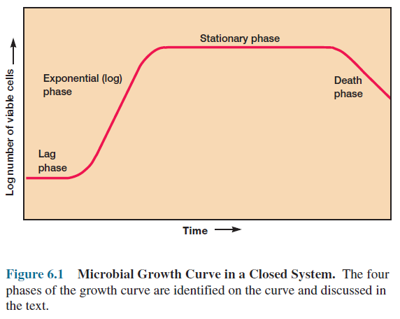
Population growth is studied by analyzing the growth curve of a microbial culture. When microorganisms are cultivated in liquid medium, they usually are grown in a batch culture or closed system— that is, they are incubated in a closed culture vessel with a single batch of medium. Because no fresh medium is provided during incubation, nutrient concentrations decline and concentrations of wastes increase. The growth of microorganisms reproducing by binary fission can be plotted as the logarithm of the number of viable cells versus the incubation time. The resulting curve has four distinct phases (figure 6.1).

Microbial Growth Curve


Lag Phase

When microorganisms are introduced into fresh culture medium, usually no immediate increase in cell number occurs, and therefore this period is called the lag phase. Although cell division does not take place right away and there is no net increase in mass, the cell is synthesizing new components. A lag phase prior to the start of cell division can be necessary for a variety of reasons.

The cells may be old and depleted of ATP, essential cofactors, and ribosomes; these must be synthesized before growth can begin. The medium may be different from the one the microorganism was growing in previously. Here new enzymes would be needed to use different nutrients. Possibly the microorganisms have been injured and require time to recover. Whatever the causes, eventually the cells retool, replicate their DNA, begin to increase in mass, and finally divide.

The lag phase varies considerably in length with the condition of the microorganisms and the nature of the medium. This phase may be quite long if the inoculum is from an old culture or one that has been refrigerated. Inoculation of a culture into a chemically different medium also results in a longer lag phase.

Exponential Phase

During the exponential or log phase, microorganisms are growing and dividing at the maximal rate possible given their genetic potential, the nature of the medium, and the conditions under which they are growing. Their rate of growth is constant during the exponential phase; that is, the microorganisms are dividing and doubling in number at regular intervals. Because each individual divides at a slightly different moment, the growth curve rises smoothly rather than in discrete jumps (figure 6.1). The population is most uniform in terms of chemical and physiological properties during this phase; therefore exponential phase cultures are usually used in biochemical and physiological studies.

Exponential growth is balanced growth. That is, all cellular constituents are manufactured at constant rates relative to each other. If nutrient levels or other environmental conditions change, unbalanced growth results. This is growth during which the rates of synthesis of cell components vary relative to one another until a new balanced state is reached. This response is readily observed in a shift-up experiment in which bacteria are transferred from a nutritionally poor medium to a richer one. The cells first construct new ribosomes to enhance their capacity for protein synthesis. This is followed by increases in protein and DNA synthesis.

Finally, the expected rise in reproductive rate takes place. Unbalanced growth also results when a bacterial population is shifted down from a rich medium to a poor one. The organisms may previously have been able to obtain many cell components directly from the medium. When shifted to a nutritionally inadequate medium, they need time to make the enzymes required for the biosynthesis of unavailable nutrients.

Consequently cell division and DNA replication continue after the shift-down, but net protein and RNA synthesis slow. The cells become smaller and reorganize themselves metabolically until they are able to grow again. Then balanced growth is resumed and the culture enters the exponential phase.

These shift-up and shift-down experiments demonstrate that microbial growth is under precise, coordinated control and responds quickly to changes in environmental conditions.

Nutrient Concentration and Growth


When microbial growth is limited by the low concentration of a required nutrient, the final net growth or yield of cells increases with the initial amount of the limiting nutrient present (figure 6.2a). This is the basis of microbiological assays for vitamins and other growth factors. The rate of growth also increases with nutrient concentration (figure 6.2b), but in a hyperbolic manner much like that seen with many enzymes (see figure 8.17).

The shape of the curve seems to reflect the rate of nutrient uptake by microbial transport proteins. At sufficiently high nutrient levels the transport systems are saturated, and the growth rate does not rise further with increasing nutrient concentration.

Stationary Phase

Eventually population growth ceases and the growth curve becomes horizontal (figure 6.1). This stationary phase usually is attained by bacteria at a population level of around 109 cells per ml. Other microorganisms normally do not reach such high population densities, protozoan and algal cultures often having maximum concentrations of about 106 cells per ml. Of course final population size depends on nutrient availability and other factors, as well as the type of microorganism being cultured. In the stationary phase the total number of viable microorganisms remains constant. This may result from a balance between cell division and cell death, or the population may simply cease to divide though remaining metabolically active.

Microbial populations enter the stationary phase for several reasons. One obvious factor is nutrient limitation; if an essential nutrient is severely depleted, population growth will slow. Aerobic organisms often are limited by O2 availability. Oxygen is not very soluble and may be depleted so quickly that only the surface of a culture will have an O2 concentration adequate for growth.

The cells beneath the surface will not be able to grow unless the culture is shaken or aerated in another way. Population growth also may cease due to the accumulation of toxic waste products.

This factor seems to limit the growth of many anaerobic cultures (cultures growing in the absence of O2). For example, streptococci can produce so much lactic acid and other organic acids from sugar fermentation that their medium becomes acidic and growth is inhibited. Streptococcal cultures also can enter the stationary phase due to depletion of their sugar supply. Finally, there is some evidence that growth may cease when a critical population level is reached. Thus entrance into the stationary phase may result from several factors operating in concert.

As we have seen, bacteria in a batch culture may enter stationary phase in response to starvation. This probably often occurs in nature as well because many environments have quite low nutrient levels.

Starvation can be a positive experience for bacteria. Many do not respond with obvious morphological changes such as endospore formation, but only decrease somewhat in overall size, often accompanied by protoplast shrinkage and nucleoid condensation. The more important changes are in gene expression and physiology.

Starving bacteria frequently produce a variety of starvation proteins, which make the cell much more resistant to damage in a variety of ways. They increase peptidoglycan cross-linking and cell wall strength. The Dps (DNA-binding protein from starved cells) protein protects DNA. Chaperones prevent protein denaturation and renature damaged proteins. As a result of these and many other mechanisms, the starved cells become harder to kill and more resistant to starvation itself, damaging temperature changes, oxidative and osmotic damage, and toxic chemicals such as chlorine.

These changes are so effective that some bacteria can survive starvation for years. Clearly, these considerations are of great practical importance in medical and industrial microbiology. There is even evidence that Salmonella typhimurium and some other bacterial pathogens become more virulent when starved.

Death Phase or Phase of Decline

Detrimental environmental changes like nutrient deprivation and the buildup of toxic wastes lead to the decline in the number of viable cells characteristic of the death phase. The death of a microbial population, like its growth during the exponential phase, is usually logarithmic (that is, a constant proportion of cells dies every hour). This pattern in viable cell count holds even when the total cell number remains constant because the cells simply fail to lyse after dying. Often the only way of deciding whether a bacterial cell is viable is by incubating it in fresh medium; if it does not grow and reproduce, it is assumed to be dead. That is, death is defined to be the irreversible loss of the ability to reproduce.

Although most of a microbial population usually dies in a logarithmic fashion, the death rate may decrease after the population has been drastically reduced. This is due to the extended survival of particularly resistant cells. For this and other reasons, the death phase curve may be complex.

The Mathematics of  Microbial Growth

Knowledge of microbial growth rates during the exponential phase is indispensable to microbiologists. Growth rate studies contribute to basic physiological and ecological research and the solution of applied problems in industry. Therefore the quantitative aspects of exponential phase growth will be discussed.

Nutrient Concentration and Growth


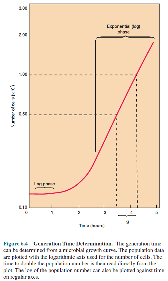
During the exponential phase each microorganism is dividing at constant intervals. Thus the population will double in number during a specific length of time called the generation time or doubling time. This situation can be illustrated with a simple example.

Suppose that a culture tube is inoculated with one cell that divides every 20 minutes (table 6.1). The population will be 2 cells after 20 minutes, 4 cells after 40 minutes, and so forth. Because the population is doubling every generation, the increase in population is always 2n where n is the number of generations. The resulting population increase is exponential or logarithmic (figure 6.3).

Bacterial generation time


Exponential Microbial Growth


Generation times vary markedly with the species of microorganism and environmental conditions. They range from less than 10 minutes (0.17 hours) for a few bacteria to several days with some eucaryotic microorganisms (table 6.2). Generation times in nature are usually much longer than in culture.

Measurement of Microbial Growth

There are many ways to measure microbial growth to determine growth rates and generation times. Either population mass or number may be followed because growth leads to increases in both. The most commonly employed techniques for growth measurement are examined briefly and the advantages and disadvantages of each noted. No single technique is always best; the most appropriate approach will depend on the experimental situation.

Bacterial Generation Time Determination


Measurement of Microbial Cell Numbers

The most obvious way to determine microbial numbers is through direct counting. Using a counting chamber is easy, inexpensive, and relatively quick; it also gives information about the size and morphology of microorganisms. Petroff-Hausser counting chambers can be used for counting procaryotes; hemocytometers can be used for both procaryotes and eucaryotes.

Generation Times for Selected Microorganisms


Procaryotes are more easily counted in these chambers if they are stained, or when a phase-contrast or a fluorescence microscope is employed. These specially designed slides have chambers of known depth with an etched grid on the chamber bottom (figure 6.5). The number of microorganisms in a sample can be calculated by taking into account the chamber’s volume and any sample dilutions required. There are some disadvantages to the technique. The microbial population must be fairly large for accuracy because such a small volume is sampled. It is also difficult to distinguish between living and dead cells in counting chambers without special techniques.

Petroff-Hausser Counting Chamber


Larger microorganisms such as protozoa, algae, and nonfilamentous yeasts can be directly counted with electronic counters such as the Coulter Counter. The microbial suspension is forced through a small hole or orifice. An electrical current flows through the hole, and electrodes placed on both sides of the orifice measure its electrical resistance. Every time a microbial cell passes through the orifice, electrical resistance increases (or the conductivity drops) and the cell is counted. The Coulter Counter gives accurate results with larger cells and is extensively used in hospital laboratories to count red and white blood cells. It is not as useful in counting bacteria because of interference by small debris particles, the formation of filaments, and other problems.

Counting chambers and electronic counters yield counts of all cells, whether alive or dead. There are also several viable counting techniques, procedures specific for cells able to grow and reproduce. In most viable counting procedures, a diluted sample of bacteria or other microorganisms is dispersed over a solid agar surface. Each microorganism or group of microorganisms develops into a distinct colony. The original number of viable microorganisms in the sample can be calculated from the number of colonies formed and the sample dilution. For example, if 1.0 ml of a 1X10⁻⁶ dilution yielded 150 colonies, the original sample contained around 1.5X 10⁸ cells per ml. Usually the count is made more accurate by use of a special colony counter.

In this way the spread-plate and pour-plate techniques may be used to find the number of microorganisms in a sample.

Plating techniques are simple, sensitive, and widely used for viable counts of bacteria and other microorganisms in samples of food, water, and soil. Several problems, however, can lead to inaccurate counts. Low counts will result if clumps of cells are not broken up and the microorganisms well dispersed. Because it is not possible to be absolutely certain that each colony arose from an individual cell, the results are often expressed in terms of colony forming units (CFU) rather than the number of microorganisms.

The samples should yield between 30 and 300 colonies for best results. Of course the counts will also be low if the agar medium employed cannot support growth of all the viable microorganisms present.

The hot agar used in the pour-plate technique may injure or kill sensitive cells; thus spread plates sometimes give higher counts than pour plates.

Membrane Filtration Procedure to count Colony


Microbial numbers are frequently determined from counts of colonies growing on special membrane filters having pores small enough to trap bacteria. In the membrane filter technique, a sample is drawn through a special membrane filter (figure 6.6). The filter is then placed on an agar medium or on a pad soaked with liquid media and incubated until each cell forms a separate colony. A colony count gives the number of microorganisms in the filtered sample, and special media can be used to select for specific microorganisms (figure 6.7). This technique is especially useful in analyzing aquatic samples.

Colonies on Membrane Filters


Membrane filters also are used to count bacteria directly. The sample is first filtered through a black polycarbonate membrane filter to provide a good background for observing fluorescent objects.

The bacteria then are stained with a fluorescent dye such as acridine orange or DAPI and observed microscopically. Acridine orange–stained microorganisms glow orange or green and are easily counted with an epifluorescence microscope. Usually the counts obtained with this approach are much higher than those from culture techniques because some of the bacteria are dead. Commercial kits that use fluorescent reagents to stain live and dead cells differently are now available. This makes it possible to directly count the number of live and dead microorganisms in a sample (see figure 2.13d).

Measurement of Microbial Cell Mass

Increases in the total cell mass, as well as in cell numbers, accompany population growth. Therefore techniques for measuring changes in cell mass can be used in following growth. The most direct approach is the determination of microbial dry weight.

Cells growing in liquid medium are collected by centrifugation, washed, dried in an oven, and weighed. This is an especially useful technique for measuring the growth of fungi. It is time consuming, however, and not very sensitive. Because bacteria weigh so little, it may be necessary to centrifuge several hundred milliliters of culture to collect a sufficient quantity.

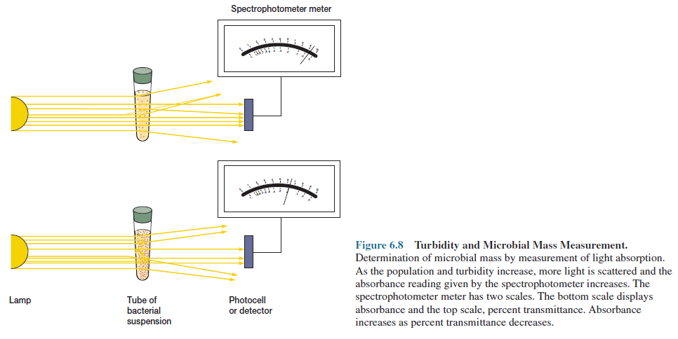
More rapid, sensitive techniques depend on the fact that microbial cells scatter light striking them. Because microbial cells in a population are of roughly constant size, the amount of scattering is directly proportional to the biomass of cells present and indirectly related to cell number. When the concentration of bacteria reaches about 10 million cells (107) per ml, the medium appears slightly cloudy or turbid.

Further increases in concentration result in greater turbidity and less light is transmitted through the medium. The extent of light scattering can be measured by a spectrophotometer and is almost linearly related to bacterial concentration at low absorbance levels (figure 6.8). Thus population growth can be easily measured spectrophotometrically as long as the population is high enough to give detectable turbidity.

Turbidity and Microbial Mass Measurement


If the amount of a substance in each cell is constant, the total quantity of that cell constituent is directly related to the total microbial cell mass. For example, a sample of washed cells collected from a known volume of medium can be analyzed for total protein or nitrogen. An increase in the microbial population will be reflected in higher total protein levels. Similarly, chlorophyll determinations can be used to measure algal populations, and the quantity of ATP can be used to estimate the amount of living microbial mass.

The Continuous Culture of Microorganisms

Up to this point the focus has been on closed systems called batch cultures in which nutrient supplies are not renewed nor wastes removed.

Exponential growth lasts for only a few generations and soon the stationary phase is reached. However, it is possible to grow microorganisms in an open system, a system with constant environmental conditions maintained through continual provision of nutrients and removal of wastes.

These conditions are met in the laboratory by a continuous culture system. A microbial population can be maintained in the exponential growth phase and at a constant biomass concentration for extended periods in a continuous culture system.

The Chemostat Continuous Culture System

Two major types of continuous culture systems commonly are used: (1) chemostats and (2) turbidostats.

Continuous Culture System: The Chemostat


A chemostat is constructed so that sterile medium is fed into the culture vessel at the same rate as the media containing microorganisms is removed (figure 6.9). The culture medium for a chemostat possesses an essential nutrient (e.g., an amino acid) in limiting quantities. Because of the presence of a limiting nutrient, the growth rate is determined by the rate at which new medium is fed into the growth chamber, and the final cell density depends on the concentration of the limiting nutrient.

The rate of nutrient exchange is expressed as the dilution rate (D), the rate at which medium flows through the culture vessel relative to the vessel volume, where f is the flow rate (ml/hr) and V is the vessel volume (ml).

D = f/V

For example, if f is 30 ml/hr and V is 100 ml, the dilution rate is 0.30 hr-1 .

Both the microbial population level and the generation time are related to the dilution rate (figure 6.10). The microbial population density remains unchanged over a wide range of dilution rates. The generation time decreases (i.e., the growth rate rises) as the dilution rate increases.

Chemostat Dilution Rate and Microbial Growth


The limiting nutrient will be almost completely depleted under these balanced conditions. If the dilution rate rises too high, the microorganisms can actually be washed out of the culture vessel before reproducing because the dilution rate is greater than the maximum growth rate. The limiting nutrient concentration rises at higher dilution rates because fewer microorganisms are present to use it.

At very low dilution rates, an increase in D causes a rise in both cell density and the growth rate. This is because of the effect of nutrient concentration on the growth rate, sometimes called the Monod relationship (figure 6.2b). Only a limited supply of nutrient is available at low dilution rates. Much of the available energy must be used for cell maintenance, not for growth and reproduction.

As the dilution rate increases, the amount of nutrients and the resulting cell density rise because energy is available for both maintenance and growth. The growth rate increases when the total available energy exceeds the maintenance energy.

The Turbidostat Continuous Culture System

The second type of continuous culture system, the turbidostat, has a photocell that measures the absorbance or turbidity of the culture in the growth vessel. The flow rate of media through the vessel is automatically regulated to maintain a predetermined turbidity or cell density. The turbidostat differs from the chemostat in several ways. The dilution rate in a turbidostat varies rather than remaining constant, and its culture medium lacks a limiting nutrient. The turbidostat operates best at high dilution rates; the chemostat is most stable and effective at lower dilution rates.

Continuous culture systems are very useful because they provide a constant supply of cells in exponential phase and growing at a known rate. They make possible the study of microbial growth at very low nutrient levels, concentrations close to those present in natural environments.

These systems are essential for research in many areas—for example, in studies on interactions between microbial species under environmental conditions resembling those in a freshwater lake or pond. Continuous systems also are used in food and industrial microbiology.

Microbial Responses to Environmental Factors


The Influence of Environmental Factors on Microbial Growth

Microorganisms must be able to respond to variations in nutrient levels, and particularly to nutrient limitation. The growth of microorganisms also is greatly affected by the chemical and physical nature of their surroundings.

An understanding of environmental influences aids in the control of microbial growth and the study of the ecological distribution of microorganisms.

The ability of some microorganisms to adapt to extreme and inhospitable environments is truly remarkable. Procaryotes are present anywhere life can exist. Many habitats in which prokaryotes thrive would kill most other organisms. Procaryotes such as Bacillus infernus even seem able to live over 1.5 miles below the Earth’s surface, without oxygen and at temperatures above 60°C.

Microorganisms that grow in such harsh conditions are often called extremophiles.

In this section we shall briefly review how some of the most important environmental factors affect microbial growth. Major emphasis will be given to solutes and water activity, pH, temperature, oxygen level, pressure, and radiation. Table 6.3 summarizes the way in which microorganisms are categorized in terms of their response to these factors.
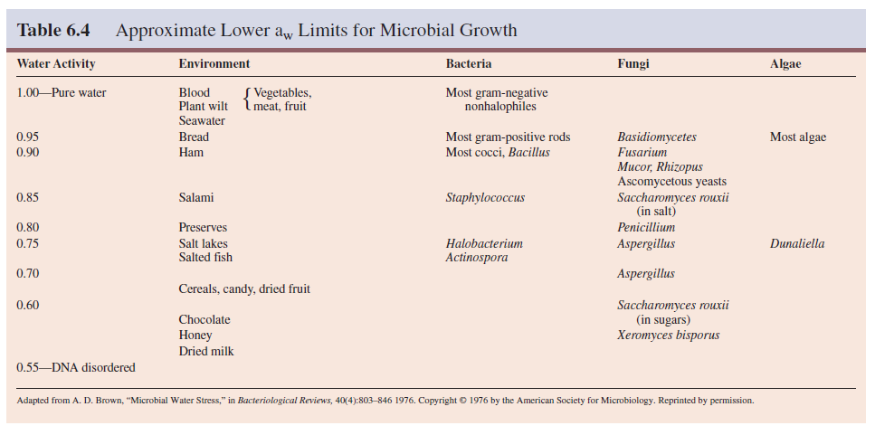
Approximate Lower aw Limits for Microbial Growth


The Influence of Solutes and Water Activity on Microbial Growth

Because a selectively permeable plasma membrane separates microorganisms from their environment, they can be affected by changes in the osmotic concentration of their surroundings. If a microorganism is placed in a hypotonic solution (one with a lower osmotic concentration), water will enter the cell and cause it to burst unless something is done to prevent the influx. The osmotic concentration of the cytoplasm can be reduced by use of inclusion bodies. Procaryotes also can contain pressure-sensitive channels that open to allow solute escape when the osmolarity of the environment becomes much lower than that of the cytoplasm.

Most bacteria, algae, and fungi have rigid cell walls that maintain the shape and integrity of the cell. When microorganisms with rigid cell walls are placed in a hypertonic environment, water leaves and the plasma membrane shrinks away from the wall, a process known as plasmolysis. This dehydrates the cell and may damage the plasma membrane; the cell usually becomes metabolically inactive and ceases to grow.

Many microorganisms keep the osmotic concentration of their protoplasm somewhat above that of the habitat by the use of compatible solutes, so that the plasma membrane is always pressed firmly against their cell wall. Compatible solutes are solutes that are compatible with metabolism and growth when at high intracellular concentrations.

Most procaryotes increase their internal osmotic concentration in a hypertonic environment through the synthesis or uptake of choline, betaine, proline, glutamic acid, and other amino acids; elevated levels of potassium ions are also involved to some extent. Algae and fungi employ sucrose and polyols—for example, arabitol, glycerol, and mannitol— for the same purpose. Polyols and amino acids are ideal solutes for this function because they normally do not disrupt enzyme structure and function. 

A few procaryotes like Halobacterium salinarium raise their osmotic concentration with potassium ions (sodium ions are also elevated but not as much as potassium). Halobacterium’s enzymes have been altered so that they actually require high salt concentrations for normal activity. Since protozoa do not have a cell wall, they must use contractile vacuoles to eliminate excess water when living in hypotonic environments.

The amount of water available to microorganisms can be reduced by interaction with solute molecules (the osmotic effect) or by adsorption to the surfaces of solids (the matric effect). Because the osmotic concentration of a habitat has such profound effects on microorganisms, it is useful to be able to express quantitatively the degree of water availability.

Microbiologists generally use water activity (aw) for this purpose (water availability also may be expressed as water potential, which is related to aw). The water activity of a solution is 1/100 the relative humidity of the solution (when expressed as a percent). It is also equivalent to the ratio of the solution’s vapor pressure (Psoln) to that of pure water (Pwater).

Aw= Psoln/Pwater

The water activity of a solution or solid can be determined by sealing it in a chamber and measuring the relative humidity after the system has come to equilibrium. Suppose after a sample is treated in this way, the air above it is 95% saturated—that is, the air contains 95% of the moisture it would have when equilibrated at the same temperature with a sample of pure water. The relative humidity would be 95% and the sample’s water activity, 0.95.

Water activity is inversely related to osmotic pressure; if a solution has high osmotic pressure, its aw is low.

Microorganisms differ greatly in their ability to adapt to habitats with low water activity (table 6.4). A microorganism must expend extra effort to grow in a habitat with a low aw value because it must maintain a high internal solute concentration to retain water. Some microorganisms can do this and are osmotolerant; they will grow over wide ranges of water activity or osmotic concentration. 

For example, Staphylococcus aureus can be cultured in media containing any sodium chloride concentration up to about 3 M. It is well adapted for growth on the skin. The yeast Saccharomyces rouxii will grow in sugar solutions with aw values as low as 0.6. The alga Dunaliella viridis tolerates sodium chloride concentrations from 1.7 M to a saturated solution.
Although a few microorganisms are truly osmotolerant, most only grow well at water activities around 0.98 (the approximate aw for seawater) or higher. This is why drying food or adding large quantities of salt and sugar is so effective in preventing food spoilage. As table 6.4 shows, many fungi are osmotolerant and thus particularly important in the spoilage of salted or dried foods.

Halophiles have adapted so completely to hypertonic, saline conditions that they require high levels of sodium chloride to grow, concentrations between about 2.8 M and saturation (about 6.2 M) for extreme halophilic bacteria. The archaeon Halobacterium can be isolated from the Dead Sea (a salt lake between Israel and Jordan and the lowest lake in the world), the Great Salt Lake in Utah, and other aquatic habitats with salt concentrations approaching saturation.

Halobacterium and other extremely halophilic bacteria have significantly modified the structure of their proteins and membranes rather than simply increasing the intracellular concentrations of solutes, the approach used by most osmotolerant microorganisms. These extreme halophiles accumulate enormous quantities of potassium in order to remain hypertonic to their environment; the internal potassium concentration may reach 4 to 7 M. The enzymes, ribosomes, and transport proteins of these bacteria require high levels of potassium for stability and activity. 

In addition, the plasma membrane and cell wall of Halobacterium are stabilized by high concentrations of sodium ion. If the sodium concentration decreases too much, the wall and plasma membrane literally disintegrate. Extreme halophilic bacteria have successfully adapted to environmental conditions that would destroy most organisms. In the process they have become so specialized that they have lost ecological flexibility and can prosper only in a few extreme habitats.

The Influence of pH on Microbial Growth

pH is a measure of the hydrogen ion activity of a solution and is defined as the negative logarithm of the hydrogen ion concentration (expressed in terms of molarity).

pH = -log [H+] = log(1/[H+])

The pH scale extends from pH 0.0 (1.0 M H+) to pH 14.0 (1.0 X 10⁻¹⁴ M H+), and each pH unit represents a tenfold change in hydrogen ion concentration. Figure 6.11 shows that the habitats in which microorganisms grow vary widely—from pH 1 to 2 at the acid end to alkaline lakes and soil that may have pH values between 9 and 10.

pH Scale


It is not surprising that pH dramatically affects microbial growth. Each species has a definite pH growth range and pH growth optimum. Acidophiles have their growth optimum between pH 0 and 5.5; neutrophiles, between pH 5.5 and 8.0; and alkalophiles prefer the pH range of 8.5 to 11.5. Extreme alkalophiles have growth optima at pH 10 or higher. In general, different microbial groups have characteristic pH preferences. 

Most bacteria and protozoa are neutrophiles. Most fungi prefer slightly acid surroundings, about pH 4 to 6; algae also seem to favor slight acidity. There are many exceptions to these generalizations. For example, the alga Cyanidium caldarium and the archaeon Sulfolobus acidocaldarius are common inhabitants of acidic hot springs; both grow well around pH 1 to 3 and at high temperatures.

The Archaea Ferroplasma acidarmanus and Picrophilus oshimae can actually grow at pH 0, or very close to it.

Although microorganisms will often grow over wide ranges of pH and far from their optima, there are limits to their tolerance.

Drastic variations in cytoplasmic pH can harm microorganisms by disrupting the plasma membrane or inhibiting the activity of enzymes and membrane transport proteins. Procaryotes die if the internal pH drops much below 5.0 to 5.5. Changes in the external pH also might alter the ionization of nutrient molecules and thus reduce their availability to the organism.

Several mechanisms for the maintenance of a neutral cytoplasmic pH have been proposed. The plasma membrane may be relatively impermeable to protons. Neutrophiles appear to exchange potassium for protons using an antiport transport system. Extreme alkalophiles like Bacillus alcalophilus maintain their internal pH closer to neutrality by exchanging internal sodium ions for external protons. Internal buffering also may contribute to pH homeostasis.

Microorganisms often must adapt to environmental pH changes to survive. In bacteria, potassium/proton and sodium/proton antiport systems probably correct small variations in pH. If the pH becomes too acidic, other mechanisms come into play. When the pH drops below about 5.5 to 6.0, Salmonella typhimurium and E. coli synthesize an array of new proteins as part of what has been called their acidic tolerance response. 

A proton-translocating ATPase contributes to this protective response, either by making more ATP or by pumping protons out of the cell. If the external pH decreases to 4.5 or lower, chaperones such as acid shock proteins and heat shock proteins are synthesized. Presumably these prevent the acid denaturation of proteins and aid in the refolding of denatured proteins.

Microorganisms frequently change the pH of their own habitat by producing acidic or basic metabolic waste products. Fermentative microorganisms form organic acids from carbohydrates, whereas chemolithotrophs like Thiobacillus oxidize reduced sulfur components to sulfuric acid. Other microorganisms make their environment more alkaline by generating ammonia through amino acid degradation.

Buffers often are included in media to prevent growth inhibition by large pH changes. Phosphate is a commonly used buffer and a good example of buffering by a weak acid (H2PO4) and its conjugate base (HPO4 2–).


If protons are added to the mixture, they combine with the salt form to yield a weak acid. An increase in alkalinity is resisted because the weak acid will neutralize hydroxyl ions through proton donation to give water. Peptides and amino acids in complex media also have a strong buffering effect.

The Influence of Temperature on Microbial Growth

Environmental temperature profoundly affects microorganisms, like all other organisms. Indeed, microorganisms are particularly susceptible because they are usually unicellular and their temperature varies with that of the external environment. For these reasons, microbial cell temperature directly reflects that of the cell’s surroundings. A most important factor influencing the effect of temperature on growth is the temperature sensitivity of enzyme catalyzed reactions.

At low temperatures a temperature rise increases the growth rate because the velocity of an enzyme-catalyzed reaction, like that of any chemical reaction, will roughly double for every 10°C rise in temperature. Because the rate of each reaction increases, metabolism as a whole is more active at higher temperatures, and the microorganism grows faster.

Beyond a certain point further increases actually slow growth, and sufficiently high temperatures are lethal. High temperatures damage microorganisms by denaturing enzymes, transport carriers, and other proteins. Microbial membranes are also disrupted by temperature extremes; the lipid bilayer simply melts and disintegrates.

Thus, although functional enzymes operate more rapidly at higher temperatures, the microorganism may be damaged to such an extent that growth is inhibited because the damage cannot be repaired. At very low temperatures, membranes solidify and enzymes don’t work rapidly. In summary, when organisms are above the optimum temperature, both function and cell structures are affected. If temperatures are very low, function is affected but not necessarily cell chemical composition and structure.

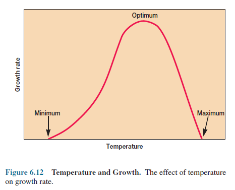
Because of these opposing temperature influences, microbial growth has fairly characteristic temperature dependence with distinct cardinal temperatures—minimum, optimum, and maximum growth temperatures (figure 6.12). Although the shape of the temperature dependence curve can vary, the temperature optimum is always closer to the maximum than to the minimum.

Temperature and Growth

The cardinal temperatures for a particular species are not rigidly fixed but often depend to some extent on other environmental factors such as pH and the available nutrients. For example, Crithidia fasciculata, a flagellated protozoan living in the gut of mosquitos, will grow in a simple medium at 22 to 27°C. However, it cannot be cultured at 33 to 34°C without the addition of extra metals, amino acids, vitamins, and lipids.

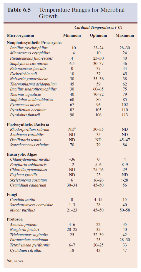
The cardinal temperatures vary greatly between microorganisms (table 6.5). Optima normally range from 0°C to as high as 75°C, whereas microbial growth occurs at temperatures extending from -20°C to over 100°C. The major factor determining this growth range seems to be water. Even at the most extreme temperatures, microorganisms need liquid water to grow. 

The growth temperature range for a particular microorganism usually spans about 30 degrees. Some species (e.g., Neisseria gonorrhoeae) have a small range; others, like Enterococcus faecalis, will grow over a wide range of temperatures. The major microbial groups differ from one another regarding their maximum growth temperature.

The upper limit for protozoa is around 50°C. Some algae and fungi can grow at temperatures as high as 55 to 60°C. Procaryotes have been found growing at or close to 100°C, the boiling point of water at sea level (see figure 20.8). Recently strains growing at even higher temperatures have been discovered (Box 6.1). Clearly, prokaryotic organisms can grow at much higher temperatures than eucaryotes.

It has been suggested that eucaryotes are not able to manufacture organellar membranes that are stable and functional at temperatures above 60°C. The photosynthetic apparatus also appears to be relatively unstable because photosynthetic organisms are not found growing at very high temperatures.

Life above 100°C


Microorganisms such as those in table 6.5 can be placed in one of five classes based on their temperature ranges for growth (figure 6.13).

1. Psychrophiles grow well at 0°C and have an optimum growth temperature of 15°C or lower; the maximum is around 20°C. They are readily isolated from Arctic and Antarctic habitats; because 90% of the ocean is 5°C or colder, it constitutes an enormous habitat for psychrophiles. The psychrophilic alga Chlamydomonas nivalis can actually turn a snowfield or glacier pink with its bright red spores. 

Psychrophiles are widespread among bacterial taxa and found in such genera as Pseudomonas, Vibrio, Alcaligenes, Bacillus, Arthrobacter, Moritella, Photobacterium, and Shewanella. The psychrophilic archaeon Methanogenium has recently been isolated from Ace Lake in Antarctica. Psychrophilic microorganisms have adapted to their environment in several ways. Their enzymes, transport systems, and protein synthetic mechanisms function well at low temperatures. 

Temperature Ranges for Microbial Growth


The cell membranes of psychrophilic microorganisms have high levels of unsaturated fatty acids and remain semifluid when cold. Indeed, many psychrophiles begin to leak cellular constituents at temperatures higher than 20°C because of cell membrane disruption.

2. Many species can grow at 0 to 7°C even though they have optima between 20 and 30°C, and maxima at about 35°C. These are called psychrotrophs or facultative psychrophiles. Psychrotrophic bacteria and fungi are major factors in the spoilage of refrigerated foods.

3. Mesophiles are microorganisms with growth optima around 20 to 45°C; they often have a temperature minimum of 15 to 20°C. Their maximum is about 45°C or lower. Most microorganisms probably fall within this category. Almost all human pathogens are mesophiles, as might be expected since their environment is a fairly constant 37°C.

Temperature Ranges for Microbial Growth


4. Some microorganisms are thermophiles; they can grow at temperatures of 55°C or higher. Their growth minimum is usually around 45°C and they often have optima between 55 and 65°C. The vast majority are procaryotes although a few algae and fungi are thermophilic (table 6.5). These organisms flourish in many habitats including composts, self-heating hay stacks, hot water lines, and hot springs. 

Thermophiles differ from mesophiles in having much more heat-stable enzymes and protein synthesis systems able to function at high temperatures. Their membrane lipids are also more saturated than those of mesophiles and have higher melting points; therefore thermophile membranes remain intact at higher temperatures.

5. As mentioned previously, a few thermophiles can grow at 90°C or above and some have maxima above 100°C.

Procaryotes that have growth optima between 80°C and about 113°C are called hyperthermophiles. They usually do not grow well below 55°C. Pyrococcus abyssi and Pyrodictium occultum are examples of marine hyperthermophiles found in hot areas of the seafloor.

The Influence of Oxygen Concentration on Microbial Growth

An organism able to grow in the presence of atmospheric O2 is an aerobe, whereas one that can grow in its absence is an anaerobe.

Almost all multicellular organisms are completely dependent on atmospheric O2 for growth—that is, they are obligate aerobes (table 6.3). Oxygen serves as the terminal electron acceptor for the electron- transport chain in aerobic respiration. In addition, aerobic eucaryotes employ O2 in the synthesis of sterols and unsaturated fatty acids. Facultative anaerobes do not require O2 for growth but do grow better in its presence. In the presence of oxygen they will use aerobic respiration.

Aerotolerant anaerobes such as Enterococcus faecalis simply ignore O2 and grow equally well whether it is present or not. In contrast, strict or obligate anaerobes (e.g., Bacteroides, Fusobacterium, Clostridium pasteurianum, Methanococcus) do not tolerate O2 at all and die in its presence. Aerotolerant and strict anaerobes cannot generate energy through respiration and must employ fermentation or anaerobic respiration pathways for this purpose.

Finally, there are aerobes such as Campylobacter called microaerophiles, that are damaged by the normal atmospheric level of O2 (20%) and require O2 levels below the range of 2 to 10% for growth. The nature of bacterial O2 responses can be readily determined by growing the bacteria in culture tubes filled with a solid culture medium or a special medium like thioglycollate broth, which contains a reducing agent to lower O2 levels (figure 6.14).

Oxygen and Bacterial Growth


A microbial group may show more than one type of relationship to O2. All five types are found among the procaryotes and protozoa.

Fungi are normally aerobic, but a number of species—particularly among the yeasts—are facultative anaerobes. Algae are almost always obligate aerobes. It should be noted that the ability to grow in both aerobic and anaerobic environments provides considerable flexibility and is an ecological advantage.

Although strict anaerobes are killed by O2, they may be recovered from habitats that appear to be aerobic. In such cases they associate with facultative anaerobes that use up the available O2 and thus make the growth of strict anaerobes possible. For example, the strict anaerobe Bacteroides gingivalis lives in the mouth where it grows in the anaerobic crevices around the teeth.

These different relationships with O2 appear due to several factors, including the inactivation of proteins and the effect of toxic O2 derivatives. Enzymes can be inactivated when sensitive groups like sulfhydryls are oxidized. A notable example is the nitrogen- fixation enzyme nitrogenase, which is very oxygen sensitive.

Oxygen accepts electrons and is readily reduced because its two outer orbital electrons are unpaired. Flavoproteins, several other cell constituents, and radiation promote oxygen reduction. The result is usually some combination of the reduction products superoxide radical, hydrogen peroxide, and hydroxyl radical.

O2 + e- → O2.– (superoxide radical)

O2.– + e- + 2H+ →H2O2 (hydrogen peroxide)

H2O2 + e- + H+ →H2O + OH- (hydroxyl radical)

These products of oxygen reduction are extremely toxic because they are powerful oxidizing agents and rapidly destroy cellular constituents. A microorganism must be able to protect itself against such oxygen products or it will be killed. Neutrophils and macrophages use these toxic oxygen products to destroy invading pathogens.

Many microorganisms possess enzymes that afford protection against toxic O2 products. Obligate aerobes and facultative anaerobes usually contain the enzymes superoxide dismutase (SOD) and catalase, which catalyze the destruction of superoxide radical and hydrogen peroxide, respectively. Peroxidase also can be used to destroy hydrogen peroxide.

Superoxide radical and hydrogen peroxide


Aerotolerant microorganisms may lack catalase but almost always have superoxide dismutase. The aerotolerant Lactobacillus plantarum uses manganous ions instead of superoxide dismutase to destroy the superoxide radical. All strict anaerobes lack both enzymes or have them in very low concentrations and therefore cannot tolerate O2.

Because aerobes need O2 and anaerobes are killed by it, radically different approaches must be used when growing the two types of microorganisms. When large volumes of aerobic microorganisms are cultured, either the culture vessel is shaken to aerate the medium or sterile air must be pumped through the culture vessel. Precisely the opposite problem arises with anaerobes; all O2 must be excluded. This can be accomplished in several ways.

(1) Special anaerobic media containing reducing agents such as thioglycollate or cysteine may be used. The medium is boiled during preparation to dissolve its components; boiling also drives off oxygen very effectively. The reducing agents will eliminate any dissolved O2 remaining within the medium so that anaerobes can grow beneath its surface.

Anaerobic Work Chamber and Incubator


(2) Oxygen also may be eliminated from an anaerobic system by removing air with a vacuum pump and flushing out residual O2 with nitrogen gas (figure 6.15). Often CO2 as well as nitrogen is added to the chamber since many anaerobes require a small amount of CO2 for best growth.

(3) One of the most popular ways of culturing small numbers of anaerobes is by use of a Gas-Pak jar (figure 6.16). In this procedure the environment is made anaerobic by using hydrogen and a palladium catalyst to remove O2 through the formation of water. The reducing agents in anaerobic agar also remove oxygen, as mentioned previously.

GasPak Anaerobic System


(4) Plastic bags or pouches make convenient containers when only a few samples are to be incubated anaerobically. These have a catalyst and calcium carbonate to produce an anaerobic, carbon-dioxiderich atmosphere. A special solution is added to the pouch’s reagent compartment; petri dishes or other containers are placed in the pouch; it then is clamped shut and placed in an incubator.

A laboratory may make use of all these techniques since each is best suited for different purposes.

The Influence of Pressure on Microbial Growth

Most organisms spend their lives on land or on the surface of water, always subjected to a pressure of 1 atmosphere (atm), and are never affected significantly by pressure. Yet the deep sea (ocean of 1,000 m or more in depth) is 75% of the total ocean volume. The hydrostatic pressure can reach 600 to 1,100 atm in the deep sea, while the temperature is about 2 to 3°C.

Despite these extremes, bacteria survive and adapt. Many are barotolerant: increased pressure does adversely affect them but not as much as it does nontolerant bacteria. Some bacteria in the gut of deep-sea invertebrates such as amphipods and holothurians are truly barophilic—they grow more rapidly at high pressures.

These gut bacteria may play an important role in nutrient recycling in the deep sea. One barophile has been recovered from the Mariana trench near the Philippines (depth about 10,500 m) that is actually unable to grow at pressures below about 400 to 500 atm when incubated at 2°C. Thus far, barophiles have been found among several bacterial genera (e.g., Photobacterium, Shewanella, Colwellia). Some members of the Archaea are thermobarophiles (e.g., Pyrococcus spp., Methanococcus jannaschii).

Electromagnetic Spectrum


The Influence of Pressure on Microbial Growth

Our world is bombarded with electromagnetic radiation of various types (figure 6.17). This radiation often behaves as if it were composed of waves moving through space like waves traveling on the surface of water. The distance between two wave crests or troughs is the wavelength. As the wavelength of electromagnetic radiation decreases, the energy of the radiation increases—gamma rays and X rays are much more energetic than visible light or infrared waves. Electromagnetic radiation also acts like a stream of energy packets called photons, each photon having a quantum of energy whose value will depend on the wavelength of the radiation.

Sunlight is the major source of radiation on the Earth. It includes visible light, ultraviolet (UV) radiation, infrared rays, and radio waves. Visible light is a most conspicuous and important aspect of our environment: all life is dependent on the ability of photosynthetic organisms to trap the light energy of the sun. Almost 60% of the sun’s radiation is in the infrared region rather than the visible portion of the spectrum.

Infrared is the major source of the Earth’s heat. At sea level, one finds very little ultraviolet radiation below about 290 to 300 nm. UV radiation of wavelengths shorter than 287 nm is absorbed by O2 in the Earth’s atmosphere; this process forms a layer of ozone between 25 and 30 miles above the Earth’s surface.

The ozone layer then absorbs somewhat longer UV rays and reforms O2. This elimination of UV radiation is crucial because it is quite damaging to living systems. The fairly even distribution of sunlight throughout the visible spectrum accounts for the fact that sunlight is generally “white.”

Many forms of electromagnetic radiation are very harmful to microorganisms. This is particularly true of ionizing radiation, radiation of very short wavelength or high energy, which can cause atoms to lose electrons or ionize. Two major forms of ionizing radiation are

(1) X rays, which are artificially produced, and

(2) gamma rays, which are emitted during radioisotope decay.

Low levels of ionizing radiation will produce mutations and may indirectly result in death, whereas higher levels are directly lethal.

Although microorganisms are more resistant to ionizing radiation than larger organisms, they will still be destroyed by a sufficiently large dose. Ionizing radiation can be used to sterilize items. Some procaryotes (e.g., Deinococcus radiodurans) and bacterial endospores can survive large doses of ionizing radiation.  

A variety of changes in cells are due to ionizing radiation; it breaks hydrogen bonds, oxidizes double bonds, destroys ring structures, and polymerizes some molecules. Oxygen enhances these destructive effects, probably through the generation of hydroxyl radicals (OH·). Although many types of constituents can be affected, it is reasonable to suppose that destruction of DNA is the most important cause of death.

Ultraviolet (UV) radiation, mentioned earlier, kills all kinds of microorganisms due to its short wavelength (approximately from 10 to 400 nm) and high energy. The most lethal UV radiation has a wavelength of 260 nm, the wavelength most effectively absorbed by DNA. The primary mechanism of UV damage is the formation of thymine dimers in DNA. Two adjacent thymines in a DNA strand are covalently joined to inhibit DNA replication and function.

This damage is repaired in several ways. In photoreactivation, blue light is used by a photoreactivating enzyme to split thymine dimers. A short sequence containing the thymine dimer can also be excised and replaced. This process occurs in the absence of light and is called dark reactivation. Damage also can be repaired by the recA protein in recombination repair and SOS repair. When UV exposure is too heavy, the damage is so extensive that repair is impossible.

Although very little UV radiation below 290 to 300 nm reaches the earth’s surface, near-UV radiation between 325 and 400 nm can harm microorganisms. Exposure to near-UV radiation induces tryptophan breakdown to toxic photoproducts. It appears that these toxic tryptophan photoproducts plus the near-UV radiation itself produce breaks in DNA strands. The precise mechanism is not known, although it is different from that seen with 260 nm UV.

Visible light is immensely beneficial because it is the source of energy for photosynthesis. Yet even visible light, when present in sufficient intensity, can damage or kill microbial cells.

Usually pigments called photosensitizers and O2 are required. All microorganisms possess pigments like chlorophyll, bacteriochlorophyll, cytochromes, and flavins, which can absorb light energy, become excited or activated, and act as photosensitizers.

The excited photosensitizer (P) transfers its energy to O2 generating singlet oxygen (1O2).

Generation of singlet oxygen


Singlet oxygen is a very reactive, powerful oxidizing agent that will quickly destroy a cell. It is probably the major agent employed by phagocytes to destroy engulfed bacteria.

Many microorganisms that are airborne or live on exposed surfaces use carotenoid pigments for protection against photooxidation.

Carotenoids effectively quench singlet oxygen—that is, they absorb energy from singlet oxygen and convert it back into the unexcited ground state. Both photosynthetic and non-photosynthetic microorganisms employ pigments in this way.

Microbial Growth in Natural Environments

The previous section surveyed the effects on microbial growth of individual environmental factors such as water availability, pH, and temperature. Although microbial ecology will be introduced in more detail at a later point, we will now briefly consider the effect of the environment as a whole on microbial growth.

Microbial Growth Limitation by Environmental Factors

The microbial environment is complex and constantly changing. Characteristically microorganisms in a particular location are exposed to many overlapping gradients of nutrients and various other environmental factors. This is particularly true of microorganisms growing in biofilms. Microorganisms will grow in “microenvironments” until an environmental or nutritional factor limits growth. Liebig’s law of the minimum states that the total biomass of an organism will be determined by the nutrient present in the lowest concentration relative to the organism’s requirements.

This law applies in both the laboratory (figure 6.2) and in terrestrial and aquatic environments. An increase in a limiting essential nutrient such as phosphate will result in an increase in the microbial population until some other nutrient becomes limiting. If a specific nutrient is limiting, changes in other nutrients will have no effect. 

The situation may be even more complex than this. Multiple limiting factors can influence a population over time. Furthermore, as we have seen, factors such as temperature, pH, light, and salinity influence microbial populations and limit growth. Shelford’s law of tolerance states that there are limits to environmental factors below and above which a microorganism cannot survive and grow, regardless of the nutrient supply.

This can readily be seen for temperature in figure 6.13. Each microorganism has a specific temperature range in which it can grow. The same rule applies to other factors such as pH, oxygen level, and hydrostatic pressure in the marine environment. The growth of a microorganism depends on both the nutrient supply and its tolerance of the environmental conditions. Biofilms.

Most microorganisms are confronted with deficiencies that limit their activities except when excess nutrients allow unlimited growth. Such rapid growth will quickly deplete nutrients and possibly result in the release of toxic waste products, which will limit further growth.

In response to low nutrient levels (oligotrophic environments) and intense competition, many microorganisms become more competitive in nutrient capture and exploitation of available resources.

Often the organism’s morphology will change in order to increase its surface area and ability to absorb nutrients. This can involve conversion of rod-shaped procaryotes to “mini” and “ultramicro” cells or changes in the morphology of prosthecate prokaryotes (figure 6.18), in response to starvation. Nutrient deprivation induces many other changes as discussed previously. For example, microorganisms can undergo a step-by-step shutdown of metabolism except for housekeeping maintenance genes.

Morphology and Nutrient Absorption


Many factors can alter nutrient levels in oligotrophic environments. Microorganisms may sequester critical limiting nutrients, such as iron, making them less available to competitors.

The atmosphere can contribute essential nutrients and support microbial growth. This is seen in the laboratory as well as natural environments. Airborne organic substances have been found to stimulate microbial growth in dilute media, and enrichment of growth media by airborne organic matter can allow significant populations of microorganisms to develop. Even distilled water, which already contains traces of organic matter, can absorb one carbon compounds from the atmosphere and grow microorganisms.

The presence of such airborne nutrients and microbial growth, if not detected, can affect experiments in biochemistry and molecular biology, as well as studies of microorganisms growing in oligotrophic environments.

Natural substances also can directly inhibit microbial growth and reproduction in low-nutrient environments. These agents include phenolics, tannins, ammonia, ethylene, and volatile sulfur compounds. This may be a means by which microorganisms avoid expending limited energy reserves until an adequate supply of nutrients becomes available. Such chemicals are also important in plant pathology and may aid in controlling soil-borne microbial diseases.

Counting Viable But Non-culturable Vegetative Procaryotes

In order to study the growth of natural procaryotic populations outside the laboratory, it is essential to determine the number of viable microorganisms present. For most of microbiology’s history, a viable microorganism has been defined as one that is able to grow actively, resulting in the formation of a colony or visible turbidity in a liquid medium.

John R. Postgate of the University of Sussex in England was one of the first to note that microorganisms stressed by survival in natural habitats—or in many selective laboratory media—were particularly sensitive to secondary stresses. Such stresses can produce viable microorganisms without the ability to grow on media normally used for their cultivation.

To determine the growth potential of such microorganisms, Postgate developed what is now called the Postgate Microviability Assay, in which microorganisms are cultured in a thin agar film under a coverslip. The ability of a cell to change its morphology, even if it does not grow beyond the single-cell stage, indicates that the microorganism does show “life signs.”

Since that time many workers have developed additional sensitive microscopic and isotopic procedures to evaluate the presence and significance of these viable but nonculturable bacteria in both lab and field. For example, levels of fluorescent antibody and acridine orange–stained cells often are compared with population counts obtained by the most probable number (MPN) method and plate counts using selective and nonselective media.

The release of radioactive-labeled cell materials also is used to monitor stress effects on microorganisms. Despite these advances the estimation of substrate-responsive viable cells by Postgate’s method is still important. These studies show that even when bacteria such as Escherichia coli, Vibrio cholerae, Klebsiella pneumoniae, Enterobacter aerogenes, and Enterococcus faecalis have lost their ability to grow on conventional laboratory media using standard cultural techniques, they still might be able to play a role in infectious disease.

The situation in natural environments with mixed populations is much more complex. Here, where often only 1 to 10% of observable cells are able to form colonies, the microbiologist is attempting to grow microorganisms that perhaps never have been cultured or characterized. In the future it is possible that media or proper environmental conditions for their growth will be developed.

At present, molecular techniques involving PCR amplification and small subunit ribosomal RNA analysis are increasingly used to analyze the diversity of uncultured microbial populations.

Quorum Sensing and Microbial Populations

For decades microbiologists tended to think of bacterial populations as collections of individuals growing and behaving independently.

More recently it has become clear that many bacteria can communicate with one another and behave cooperatively. A major way in which this cooperation is accomplished is by a process known as quorum sensing or autoinduction. This is a phenomenon in which bacteria monitor their own population density through sensing the levels of signal molecules, sometimes called autoinducers because they can stimulate the cell that releases them.

The concentration of these signal molecules increases along with the bacterial population until it rises to a specific threshold and signals the bacteria that the population density has reached a critical level or quorum. The bacteria then begin expressing sets of quorum-dependent genes. Quorum sensing has been found among both gram-negative and gram-positive bacteria.

Quorum sensing makes good practical sense. Take the production and release of extracellular enzymes as an example. If such enzymes were released by only a few bacteria, they would diffuse away and be rendered ineffective because of dilution.

With control by quorum sensing, the bacteria reach a high population density before they release enzymes, and as a consequence enzyme levels are concentrated enough to have significant effects.

This is an advantage within a host’s body as well as in the soil or an aquatic habitat. If a pathogen can reach high levels at a particular site before producing virulence factors and escaping into surrounding host tissues, it has a much better chance of counteracting host defenses and successfully spreading throughout the host’s body. This explains another pattern in quorum sensing. It seems to be very important in many bacteria that establish symbiotic or parasitic relationships with hosts.

Quorum sensing was first discovered in gram-negative bacteria and is best understood in these microorganisms. The most common signals in gram-negative bacteria are acyl homoserine lactones (HSLs). These are small molecules composed of a 4- to 14-carbon acyl chain attached by an amide bond to homoserine lactone (figure 6.19a). The acyl chain may have a keto group or hydroxyl group on its third carbon. 

Quorum Sensing in Gram-Negative Bacteria


Acyl HSLs diffuse into the target cell (figure 6.19b). Once they reach a sufficiently high level, acyl HSLs bind to special receptor proteins and trigger a conformational change. Usually the activated complexes act as inducers—that is, they bind to target sites on the DNA and stimulate transcription of quorum-sensitive genes. The gene needed to synthesize acyl HSL is also produced frequently, thus amplifying the effect by the production and release of more autoinducer molecules.

Many different processes are sensitive to acyl HSL signals and quorum sensing in gram-negative bacteria. Some well-studied examples are (1) bioluminescence production by Vibrio fischeri, (2) Pseudomonas aeruginosa synthesis and release of virulence factors, (3) conjugal transfer of genetic material by Agrobacterium tumefaciens, and (4) antibiotic production by Erwinia carotovora and Pseudomonas aureofaciens.

Gram-positive bacteria also regulate activities by quorum sensing, often using an oligopeptide signal. Good examples are mating in Enterococcus faecalis, competence induction in Streptococcus pneumoniae, stimulation of sporulation by Bacillus subtilis, and production of many toxins and other virulence factors by Staphylococcus aureus. Quorum sensing even stimulates the development of aerial mycelia and the production of streptomycin by Streptomyces griseus. In this case, the signal seems to be γ-butyrolactone rather than an oligopeptide.

An interesting and important function of quorum sensing is to promote the formation of mature biofilms by the pathogen Pseudomonas aeruginosa, and it may play a role in cystic fibrosis.

Biofilm formation makes sense for the pathogen because biofilms protect against antibiotics and detergents. Quorum sensing should be very effective within biofilms because there will be less dilution and acyl HSL levels will increase rapidly. Under such circumstances, two different bacteria might stimulate each other by releasing similar signals; this appears to be the case in biofilms containing the pathogens P. aeruginosa and Burkholderia cepacia.

Quorum sensing is an example of what might be called multicellular behavior in that many individual cells communicate and coordinate their activities to act as a unit. Other examples of such complex behavior is pattern formation in colonies and fruiting body formation in the myxobacteria.